

| Wally Welker, Roger Reep, J.I. Johnson |
| This
project has provided the first complete sectioning of whole brains
of the Florida manatee (Trichechus manatus laterostris).
Nine brains were cut in different planes of section and stained
for nerve cells and fibers. Systematic microscopic study of the
neuronal architecture of these specimens has provided us with a
comprehensive overview of how the manatee's internal brain structures
are organized. Alternate sections, stained for cell bodies and for
myelinated fibers (in transverse, sagittal and horizontal planes)
reveal details about the relative sizes, degree of complexity, and
spatial distribution of the many cellular groups and fiber tracts.
All of the brain structures found in manatees are common to most
mammals. But in the manatee many of these nuclei and tracts are
differentially developed.
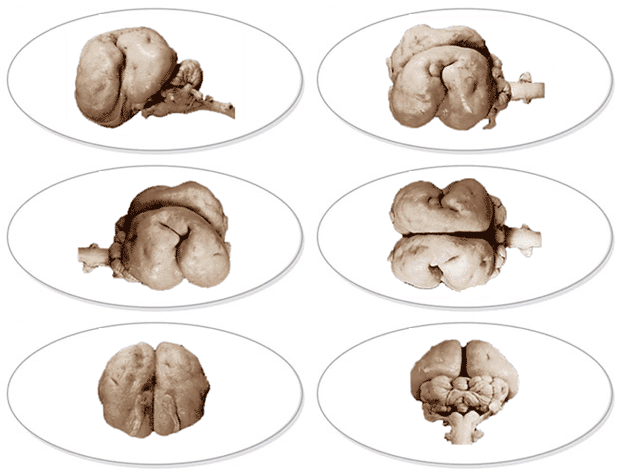
Macroscopic and microscopic study of our anatomical specimens reveal several distinguishing features of the brain of manatees: Large, lobulated somatic sensory nuclei of the brainstem that process information from the manatee's mobile browsing mouth parts (trigeminal nuclei), forelimb (cuneate nucleus) and the fluke (Bischoff's nucleus); Somatic sensory regions from the face are also relatively enlarged in the thalamus as well as the cerebral cortex. Facial motor nuclei in the medulla that produce the prominent orofacial behaviors of the proboscis, lips and vibrissae are relatively large. Particularly large are all the sensory brain structures of the auditory system, which indicate that hearing is well developed in manatees. This hypertrophy is seen in the massive auditory nuclei of the brainstem and thalamus, especially the cochlear nuclei, nuclei of the lateral lemniscus, and medial geniculate nucleus. The region of cerebral cortex estimated to be auditory in function is also relatively large; Also, noticeable are the relatively small eye, optic nerve and sensory visual regions of the lateral geniculate nucleus, superior colliculus, and thalamus; Relatively small, too, are the motor nuclei that innervate eye muscles (oculomotor, trochlear and abducens nuclei); The brainstem sensory and motor nuclei associated with the animal's large tail and enlarged viscera are also more complex and enlarged; Olfactory circuits are small, but taste circits are prominent. Of special note is that the manatee's cerebral cortex is relatively smooth and unconvoluted, suggesting that this cortex is less complexly interconnected within and between cortical areas, as well as with extracortical regions of the brain. The implied relative simplicity of cortical circuits perhaps reflects the relative simplicity of the manatee's sensory-motor life-history traits Brain nuclei (amygdala,basal forebrain nuclei, hypothalamus, hippocampus) associated with socio-emotional expression are not very elaborate or enlarged in manatees. Again, such relative simplicity may reflect the relatively calm social and emotional lifestyles often noted of these mammals. These various features indicate a minimal behavioral role of vision; a perceptuo-motor emphasis on audition, and upon somato-sensation from perioral structures (expected from a browsing animal with large mobile proboscis and vibrissae) as well as from the forelimb and fluke (not expected). We are preparing a series of papers (electronic and hardcopy) which, for the first time, will provide a detailed description and portrayal of the important brain features (mentioned above) of this unique endangered marine mammal.
|
| UNIVERSITY
OF WISCONSIN |
UNIVERSITY
OF FLORIDA |
MICHIGAN
STATE UNIVERSITY |
NATIONAL
SCIENCE FOUNDATION |
| Wally Welker | Roger Reep | John Irwin Johnson | Sponsor |
Illustrations by Carol Dizack
Website Design by Mary Walsh