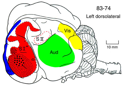
The
drawing of the manatee brain shown above indicates the sensory
and motor areas of cerebral cortex as suggested by the external
morphology as well as by the microscopic appearance of the manatee
cortex. This picture indicates the well accepted notion that
cerebral cortex is the target region where the sensory environment
is mapped in an orderly topographic fashion.
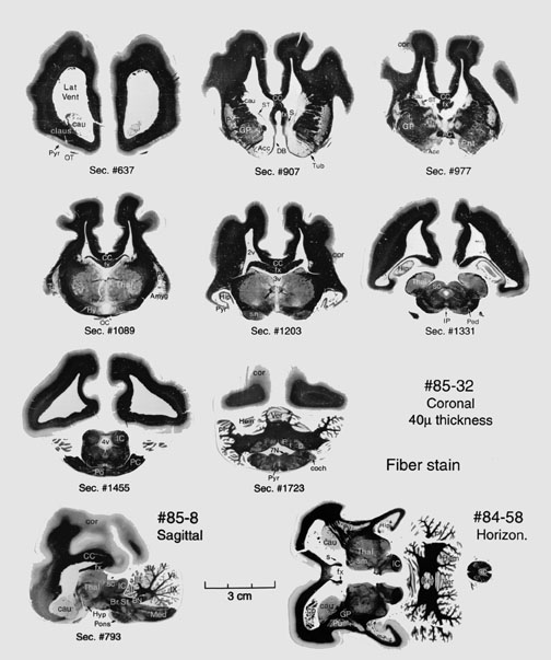
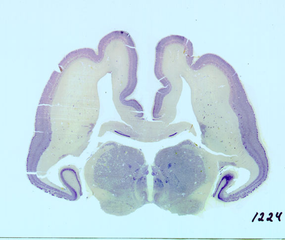
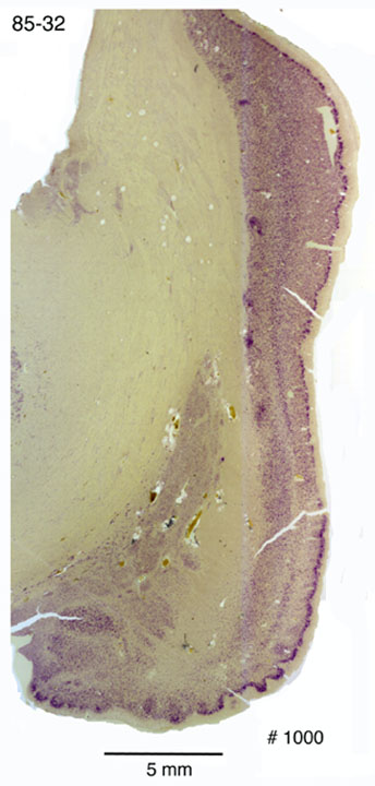
The cerebral cortex consists of collections of cellular regions arranged as a sheet which covers the entire forebrain in mammals. It is generally subdivided into two general zones: the neocortex and the paleocortex, which merge into transitional cortical zones together where they adjoin.
Neocortex
The neocortex is a seven-layered sheet of neurons which may be quite elaborately convoluted into an extensive array of gyri which may contain a large number of independently developed cortical areas. These cortical areas are richly interconnected with each other and with the thalamus, basal forebrain and even more distant brainstem targets. These connections are usually very specific and often reciprocal. Neocortex lies on the dorsal, dorsomedial, dorsolateral, frontal, caudal and ventrolateral portions of each hemisphere. The cortical regions in the two hemispheres are connected to each other via a large fiber bundle known as the corpus callosum. The major sensory inputs from the environment or the body itself project to cerebral neocortex in ways that represent the real features of the external world. Paleocortex These regions of cerebral cortex can be seen from the coronal series of sections displayed in the brain atlas and in the array of sections cut in coronal, horizontal and sagittal planes which are displayed below. These sections were all stained to show fiber stains, and cerebral cortex is revealed as the paler grey sheet overlying the forebrain. The cell bodies themselves are revealed below as purple dots in a coronal section through the center of the manatee's brain stained with thionin. |