|
J.
I. Johnson, Wally Welker and R.C. Switzer III.
Anatomy Dept. and Neuroscience Program, Michigan State Univ., East Lansing, MI 48824; Dept. of Neurophysiology, Univ. of Wisconsin-Madison, Madison, WI 53706; Neuroscience Associates, Knoxville, TN 37922. Supported
by the National Science Foundation Grant nos. BNS 9111952, IBN
9318819, BNS 899438.
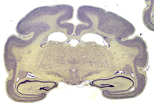
ABSTRACTOur purpose was to determine the optimal means of capturing electronic images of stained sections of mammalian brains, at both high and low magnifications(whole sections). Three methods proved optimal, depending on the size of the field to be imaged. 1) For sections or fields over 25 mm in length or width, direct scanning of the tissue in a good quality desktop scanner (e.g. the LaCie Silverscanner II), using the Transparency mode; 2) For those less than 15 mm in length or width, a microscope is used with either a film camera (to make 35 mm slides for subsequent scanning) or a digital camera (in our tests, the Kodak DCS 200)3) For those between 15 mm and 25 mm, optimal results are obtained using a camera (film or digital), our Leitz series (or their equivalents by Nikon) of "macro" (24 to 80 mm) lenses, and a specially designed, portable, slide holder and illuminator. Images obtained from the desktop scanner or the digital camera had two advantages over those captured from film and subsequently scanned: 1) much less editing was needed to produce good final results, and 2) the electronic images were immediately available, avoiding the time-consuming processes of film development and subsequent scanning. It was further determined that for our brain sections, computer image files of 150 pixels per inch, with a maximum dimension of 1050 pixels, stored as PICT files, compressed by the "high-quality" level of JPEG compression included in the PICT file creation module of the Adobe Photoshop program, proved best for economical electronic storage and transmission, for on-screen viewing, and for making good quality prints. An electronic version of this poster is in preparation for viewing on the Internet at http://www.neurophys.wisc.edu/brain/ (Supported by NSF grants BNS 9111952, IBN 9318819, and BNS 899438.) Abstract |
List of Specimens | Explore Collections | Brain Sections | Brain Evolution | Brain Development | Brain Circuitry | Brain Functions | Location and Use | Related Web Sites | Contact Us | Search MSU Database | Personnel | Home