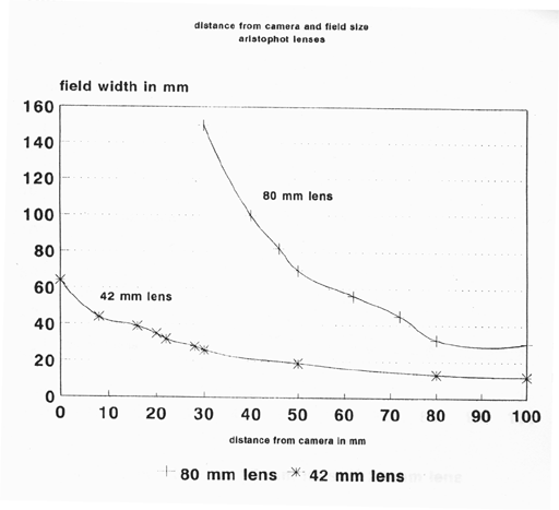
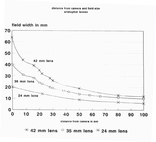
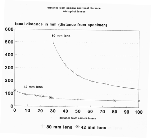
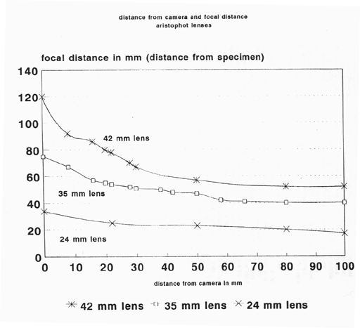
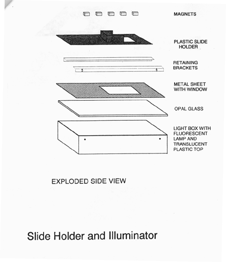
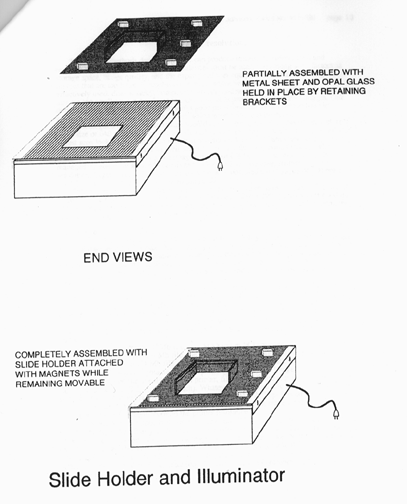
MethodsMETHODS FOR IMAGE CAPTUREFor any of the following methods, it is a good idea to include a portion of a millimeter ruler in the field, to serve as a guide to magnifications and reductions. If a large number of images are being captured at the same magnification, the ruler need appear in only a few, widely spaced, images. 1. Direct ScanningFor images larger than 15 mm in their longest dimension, our best results are obtained from scanning the section directly on a flat-bed desktop scanner (Silverscanner II from La Cie with transparency adapter). The images produced are equal in resolution to those produced via 35 mm film and scanning from 35 mm film, and the background and color fidelity is superior to those made from film intermediates. 2. Photo CDFor images smaller than 15 mm, we have photographed sections at any desired magnification, using high-quality lenses (macroscopic or microscopic) and 35 mm color film. The images on film are digitized using Kodak Photo CD processing. At this time the Kodak process yields the cheapest high-quality scanning available for large numbers of images on film. 3. Digital CameraSomewhat better, much faster, and ultimately perhaps cheaper results can be obtained by photographing specimens directly with a digital camera. The first digital camera we were able to use, the Kodak DCS 200, produced images rather better than those obtained from Photo CD. The images produced from the DCS200 camera share the advantages of those produced from large fields by the SilverScanner: clear background, good color fidelity, resolution as good as that seen in Photo CD, and little editing needed. Using this digital camera, images are obtained within 5 seconds; there is no need for film developing and waiting (up to 1 month lately) for Photo CD processing. The next camera tested, the newly available Leaf Lumina, costs about 1/4 less than the Kodak camera, captures at a higher resolution than the Kodak camera and yields noticeably better images. Capture time, however, is about 7 minutes per image. Both cameras are compatible with Nikon macro lenses, but the Leaf Lumina is not compatible for use with microscopes, while the Kodak DCS 200 produces fast, very good images from the microscope. While the cost of either of the cameras is considerable, their cost is more than made up by the time they save in editing, which is the most expensive aspect of image capture. (In our experience, images captured by digital camera require about 80% less editing time than those captured through color film.) 4. Scanning Large-Format Negative FilmWe obtained the highest resolution images by scanning black and white, large format, 4 x 5 in. negative film on the desktop scanner in Transparency mode. For our purposes, the production of a catalog of the brain collections, this is not a practical method for acquiring large numbers of images at relatively low magnification. But for high-magnification views of restricted portions of sections, this could be the most desirable means of electronic rendition of image data for particular research purposes. It may be the best means of producing detailed atlases for research use. The file size of the images (13 to 50 Mb) is not practical for use with currently generally available equipment; this will probably change in the future. We have not yet tested large-format color film. Detailed Methods1. Tabletop Scanning ProceduresIn transparency mode, we place a section on its slide, situated so that the section (not the slide) is oriented as desired relative to the vertical and horizontal margins of the transparency window on the scanner surface. We place a transparent millimeter rule somewhere near the section on the scanner surface. We scan at 800 ppi (pixels per inch), as a compromise between our scanner's default 400 ppi and its maximum 1600 ppi. The maximum resolution, for the size sections we usually use, yields a file of 18 Mb, too large to work with conveniently. The 800 resolution gives a file of 4 Mb, 400 a file of 1 Mb. The 800 resolution is also a compromise between better contrast and better detail fidelity in eventual printing (see section on Prints and Resolutions below). For a series of related sections, we find the largest section in the series that we intend to capture. We preview the section on the scanner, and create the scanning marquee so that it contains the section, a comfortable margin around the section, and a segment of the ruler showing at least 5 mm. We record the size of the marquee window (this is displayed on the scanner menu). We use this window for all subsequent sections in the series. The window can be dragged around to enclose each of the subsequent sections. This keeps all images in the series the same relative size (we keep a piece of ruler in all the images anyway, as a check on keeping all images the same relative size). We then scan the images. Some care in placing slides on the scanner surface keeps the orientation of successive images in the marquee window (hence on the computer screen) fairly constant, this saves much work later in getting images into register with one another. The images appear in the Adobe Photoshop program; we save them from there as PICT files using the High Quality JPEG level in the PICT file option within the Save menu of the Photoshop program. Photo CD and Digital Camera ProceduresUse With Microscope For images less than 5 mm in maximal width, our best results were obtained using a 35 mm film camera (for Photo CD) or the Kodak DCS 200 Digital camera, mounted on a good quality research microscope. Such microscopes have appropriate lenses, condensers and light sources to produce excellent images. Use with Macro Lenses For images more than 5 mm, but less than 15 mm in maximal width, our best results were obtained using the Leaf Lumina scanning camera with our Leitz series of macro lenses. Using the same lenses with a 35 mm film camera yielded good images for processing by Photo CD, but not as good as those obtained with the Lumina. In contrast to using a research microscope, using macro lenses requires careful attention to the light source; and to the optimal distances between the specimen, the lens, and the camera, which vary considerably with the image size. Optimal Distances The charts on the following pages show the optimal distances, empirically determined using a 35 mm film camera, for images with their maximal dimension in a range of sizes. We used Leitz-Wetzlar Summar lenses of 24, 35, 42 and 80 mm. On the charts, distance from camera refers to the length of an extension tube or extension bellows used to move the lens some distance away from the front of the camera. Focal distance refers to the distance between the lens and the specimen when the specimen is in focus. Referring first to a field size chart, it can be determined that for a specimen of 50 mm width, the 42 mm or the 80 mm lens can be used; the 42 mm lens requires 5 mm of extension tube or bellows, the 80 mm lens requires 50 mm extension. Referring next to the focal distance chart, it is seen that the 42 mm lens should be 50 mm from the specimen; the 80 mm lens should be 250 mm from the specimen. Light Source Our best results were obtained using a flat-field light source, eliminating the need for optical condensers. To provide this illumination, we devised and constructed a portable Slide Holder and Illuminator for macrophotography, illustrated on the following pages. Its basic unit is a commercially available Logan Desktop Light Box (No. 810/920, Logan Electric Spec. Mfg. Co., Chicago, IL 60622). This box has outside dimensions of 12.5 x 9.5 x 2.5 inches. It contains one fluorescent tubular lamp, color corrected to 5000xK, centered in front of a hemicylindrical reflector which reflects light through a translucent plastic top. This top is held in place by retaining brackets, which can be loosened, or removed and then replaced, to allow alternative top pieces to be inserted. There is a wire bracket on the side so that the box can be hung vertically, as well as set on a surface horizontally. Over the plastic top, and retained in the brackets, we placed a sheet of Eastman Kodak Opal Glass to maximize evenness of illumination. Over this, and also retained in the brackets we inserted a sheet of metal (galvanized steel) with a window cut out. In our most used version, the window was 4 x 6 inches. Placed on top of this, and not retained in the brackets, we placed a slide holder made of 1/8 inch Plexiglas, painted flat black. This also had a window (in the most used version this window was 3 x 4 inches). Along one of the 4 inch and one of the three inch sides of this window, another strip of black-painted plastic was fastened at right angles to the plane of the window, and a groove was cut into the window side of the strips 1 inch above the plane of the window. A third similar strip extended from the 3-inch strip down the other 4 inch side for a distance of 1.3 inches. The grooves cut in the three strips formed a slot into which could be inserted any glass slide with one dimension of 3 inches; we used this arrangement for slides of 3 x 1, 3 x 2 and 3 x 4 inches. Similar holders were constructed, with larger windows, for larger slides. Inexpensive small ceramic magnets were used to hold the plastic slide holder against the metal sheet. Magnets can be added or removed to adjust the tightness of the hold, but they always allow for gross and fine movements of the holder. These movements allow fine manual control of the placement of sections on the slides in the field of view of the camera lenses. The apparatus permits a full range of rotary and linear motions restricted to the plane of the slide, and keeps the sections firmly in place at the end of a movement. The raised slots which hold the section keep the specimen one inch above the surface of the opal glass. This insures that any specks or other defects in that surface will be out of the focal plane and will not mar the resulting pictures after photography. The apparatus can be used horizontally with an overhead camera, or vertically placed in front of a camera. It is eminently portable, and can be carried in a tote bag, backpack, or briefcase. Henry Wieferich, technician in the Psychology Department, Michigan State University, and Dr. Robert C. Switzer III of Neuroscience Associates, Knoxville, TN, provided much valuable input in the design and construction of this apparatus. Choice Of ResolutionEach of the methods described above can produce images of various sizes and resolutions. In selecting among these, balances must be achieved among such factors as storage space, image quality, and the capacity of user equipment to deal with image files. Images that are too large, in resolution or size, cannot be effectively stored, transmitted, or effectively used: disk capacity, computer memory, and retrieval time are limited. Images that are too small are not good images. Our goal is to produce good, usable images. Our definition of usable is an image that looks good on-screen, can be accommodated in generally available equipment, and from which good prints can be made. To look good on-screen, an image should have a resolution of 72 pixels per inch (ppi) and a size of 640 x 420 pixels. Images viewed at less than their true size are distorted (this is best seen looking at print on an image at different sizes in different viewing programs), and images viewed at larger than their true size are rapidly and obviously distorted ("pixelized"). Such optimally sized images are about 0.8 Mb in size, and are readily managed by generally available computers. Good prints, however, require higher resolution, larger images. Our best results have been obtained with images of 200 ppi over 8 x 10 inches (725 X 576 pixels), with image size 8 Mb. To compromise between these image sizes, our tests have shown a size of 2.1 Mb on screen, with a resolution of 150 ppi over 5 x 7 inches (1050 x 700 pixels) yields good on-screen images and good prints (after expanding the file by interpolation to 8 Mb using the standard printing software - Adobe Photoshop). Prints and ResolutionsOur findings are based on examination of high quality color prints, printed using a dye-sublimation printer (Kodak XL 7700) by DLM Imaging, Madison, Wisconsin. Images were captured from a given sample of tissue at various resolutions using two capture mechanisms: 1) scanning the tissue directly in a good quality desktop scanner (SilverScanner II from LaCie) in transparency mode, and 2) Kodak Photo CD from film images. The scanner scans at 1600, 800, and 400 (the default level) pixels per inch (ppi), as well as a number of lower resolutions (300, 150, etc.) We made scans at the three highest levels. For our size of image, about 1.5 x 2.5 inches, the highest resolution, 1600 ppi, yields files of 18 MB, 800 ppi yields 4 Mb files, and 400 ppi yields 1 Mb files. The Photo CD process yields an "image pack" from a given film image, the same image in five different resolutions. For our test we used the three highest-resolution images:
For prints on the dye sublimation printer, files of 8 Mb are required, and the printer has a resolution of 210 dpi (dots per inch). Whatever their initial size and resolution, files were resized in Adobe Photoshop to conform to this 8 Mb, 210 dpi format. The question being tested was, what difference does it make what the original resolution was, before conversion to the printer resolution? The answer obtained held for both the Photo CD and the scanned images. Starting with higher resolution files yielded prints with less contrast, but finer shadings of detail and color. With some images this is advantageous; for other images it was better to have the greater contrast and better outlines obtained from lower resolution initial files. Starting with lower resolution files produced prints which looked very similar to what is produced on-screen by applying a sharpening filter (e.g. Unsharp Mask in Photoshop) to the higher resolution image. Editing After DigitizingAfter digitizing by direct scanning, digital camera, or Photo CD, we retrieve the image, edit it (using Adobe Photoshop), and apply any desired labeling. In the editing process, contrast, color balance, background, and position of the image in the screen frame are optimized. Editing time various according to capture method and computer speed, ranging from 40 minutes when captured from film and edited with a Macintosh IIci computer, to less than 3 minutes when captured directly from tissue by a scanner or digital camera and edited with a Power Mac 8100 computer. Editing the same image with the Macintosh IIci and the Power Mac 8100 shows that the difference in computer speed cuts editing time for a particular image by more than half. Steps followed in the editing procedure: 1. Draw a rectangular marquee around a segment of the ruler containing 3, 5, or 10 mm, selecting it. Move it as close as possible to the edge of the section. Keep it visible next to the section through all the editing steps below, until the last one. If necessary, while selected, rotate it (moving it away from the section if necessary to avoid overlapping the section, but put or keep it close to the edge of the section) so that it is parallel to a horizontal edge of the screen window. Then, while still selected, copy it to the clipboard. If you should lose it in the editing process, it can be retrieved by pasting from the clipboard. 2. Lasso the section and the ruler segment, choose inverse from the Select menu, use the eyedropper tool to put the background color as the window background color (option - click on the background next to the section. Press the delete key: this will turn everything outside the lassoed window to the background color. 3. If necessary, crop the window to provide a pleasing margin around the image of the section. Show Rulers from the Windows menu, and note the length of the horizontal and vertical edges (this will be needed to crop the remaining images to the same size, even if the original image was not cropped.) 4. Resize the image, under Image Size in the Image Menu, to 1050 pixels wide and 150 ppi resolution. Then resize the Canvas, under Canvas Size in the Image Menu, to 1050 pixels wide and 650 pixels high. 5. Do any editing of Levels (contrast, command L), Color Balance (command Y), or hue/saturation (command U) [these can also be accessed under Adjust in the Image menu. 6. Lasso the section (and ruler) and move it to the center of the window. Note the ruler positions of any landmarks to be used in bringing other sections into register. To Keep sections in different files in register:
7. Type in any identification labels. The text-writing module in Photoshop allows the same text to be inserted in many successive images, without re-writing. 8. Lasso or marquee the ruler segment, rotate it if necessary to bring it parallel to the horizontal margin, then move it to a position below the desired location of the final scale bar. 9. Draw a rectangular marquee above the ruler segment, and draw a neat new ruler, using the line tool and the marquee borders and the image of the original ruler segment as guides. Then draw a rectangular marquee around the original ruler and delete it; then draw another rectangular marquee above the new drawn ruler such that it cuts into the teeth of the rule; delete it to even the ends of the teeth. Type in the scale bar label ( e.g. "5 mm"). 10. Save the completed image as High Quality JPEG PICT file. 11. Repeat the process for the rest of the images in the series. Photomatic should be able to do all of this except the cropping, lassoing and marquees. Photomatic is a program, from Daystar, which will record your actions during editing, and then replay them to work on additional images. It will faithfully repeat most editing commands, BUT NOT THOSE INVOLVING MOUSE-DRAGGING, which somewhat limits its usefulness. Digital File Compression and StorageAfter capture and editing, the resulting 2.1 Mb files are stored on disks as PICT files, using the "high-quality" level of JPEG compression available in the Photoshop PICT file saving menu. Unlike "lower quality" JPEG compressed files, our files do not undergo noticeable degradation with repeated "high quality" compression and decompression. When compressed, the image file occupies about 100 kb (0.1 Mb) of disk storage space. Thus thousands of such images can be accommodated on a single 680 Mb CD-ROM. |
List of Specimens | Explore Collections | Brain Sections | Brain Evolution | Brain Development | Brain Circuitry | Brain Functions | Location and Use | Related Web Sites | Contact Us | Search MSU Database | Personnel | Home